

When I encounter pigmentation right up against the eye, before choosing a laser wavelength I first reconfirm what this pigment actually is. Even if they look similar on the surface, lentigo and melasma differ in their cause, the depth at which they settle, and the way they are treated. Today I would like to record a case in which I carefully treated a lentigo on the left side of the eye area.
One finger's width from the eye: the location was my first concern
A woman in her mid-40s came in for a consultation about pigmentation near her left eye. The lesion itself was a lentigo with relatively well-defined borders, but because it sat very close to the eye, I had the impression from the first look that the approach would not be simple.
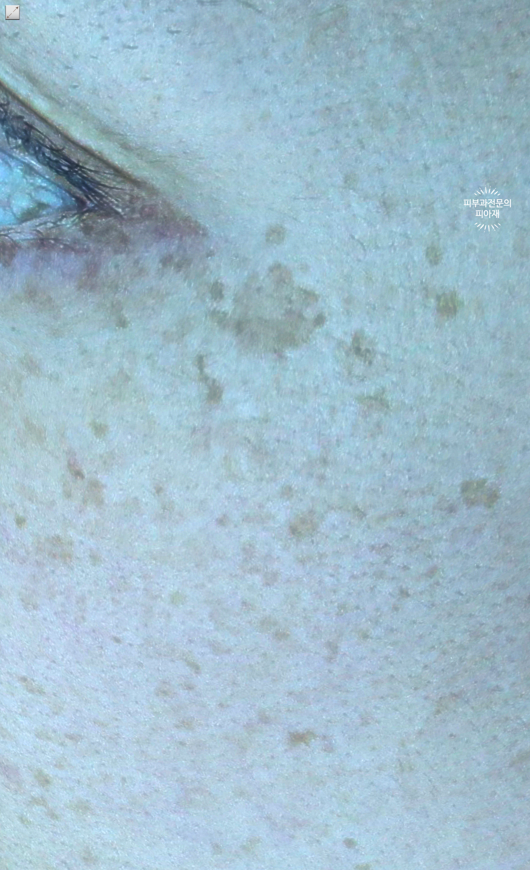
With pigmentation near the eyelid, two things need attention at the same time. One is setting the treatment area while reliably protecting the eye; the other is finely adjusting the wavelength and fluence so that excessive energy does not remain in the thin skin around the eye. So even for the same lentigo, the difficulty of treatment varies considerably depending on where it is.
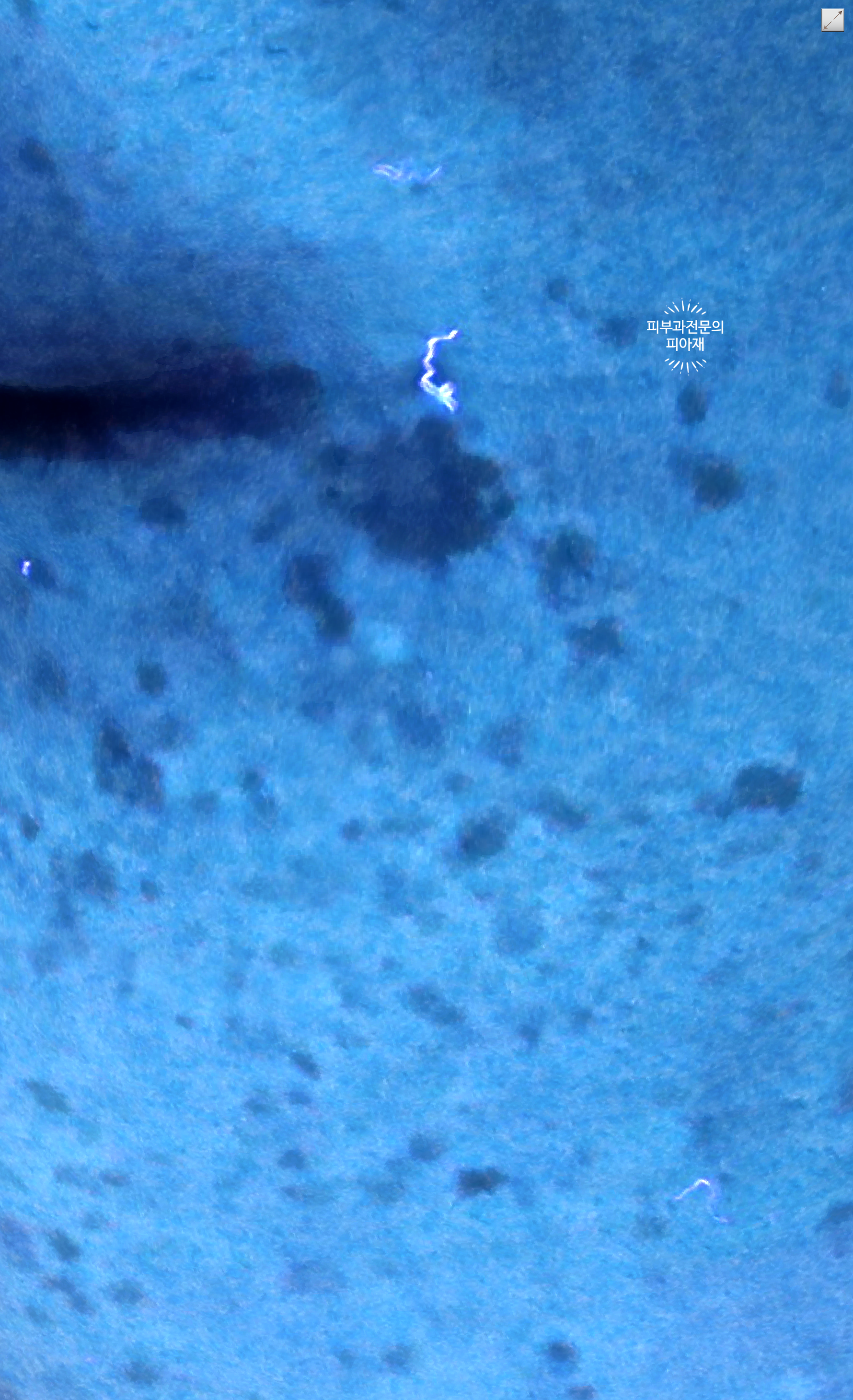
Why lentigo and melasma are not the same pigment
This is the first thing I sort out in the clinic. Looking only at the appearance of brown pigment, they are hard to tell apart, but histologically the two pigments have different characters.
| Category | Lentigo | Melasma |
|---|---|---|
| Main background | Increased melanin in the epidermal basal layer | Often involves the epidermis and dermis together |
| Borders | Relatively well defined | Blurred, as if spreading |
| Response to stimulation | Relatively predictable | Can actually darken with stimulation |
| Approach | Wavelength selected to target the lesion | Stepwise management that also considers the background |
This difference matters because it determines the treatment strategy. If an approach that should be tailored to lentigo is applied as if it were melasma, the response is slow; conversely, when melasma is pushed hard all at once as if it were lentigo, the pigment is often seen in the clinic to darken instead. I explain that lentigo is closer to a pigment you target and remove, while melasma is closer to a pigment whose background must be read as well.
The same visible color does not mean the same cause. Only once you have determined what kind of pigment it is can you decide which tool to use.
I have written in more detail about how I categorize pigmentation in How a dermatologist looks at pigmentation and Why I divide pigmentation that patients call age spots into three types. If you have any questions, feel free to leave them via Consult via chat now.
Why I chose the Reepot laser for this eye-area lentigo
In this case, I treated the pigment using the Reepot laser. For pigment with defined borders such as lentigo, I choose a wavelength that acts selectively on the melanin of the lesion, and in a sensitive area such as around the eye, I adjust the fluence so as to leave less burden on the surrounding skin.
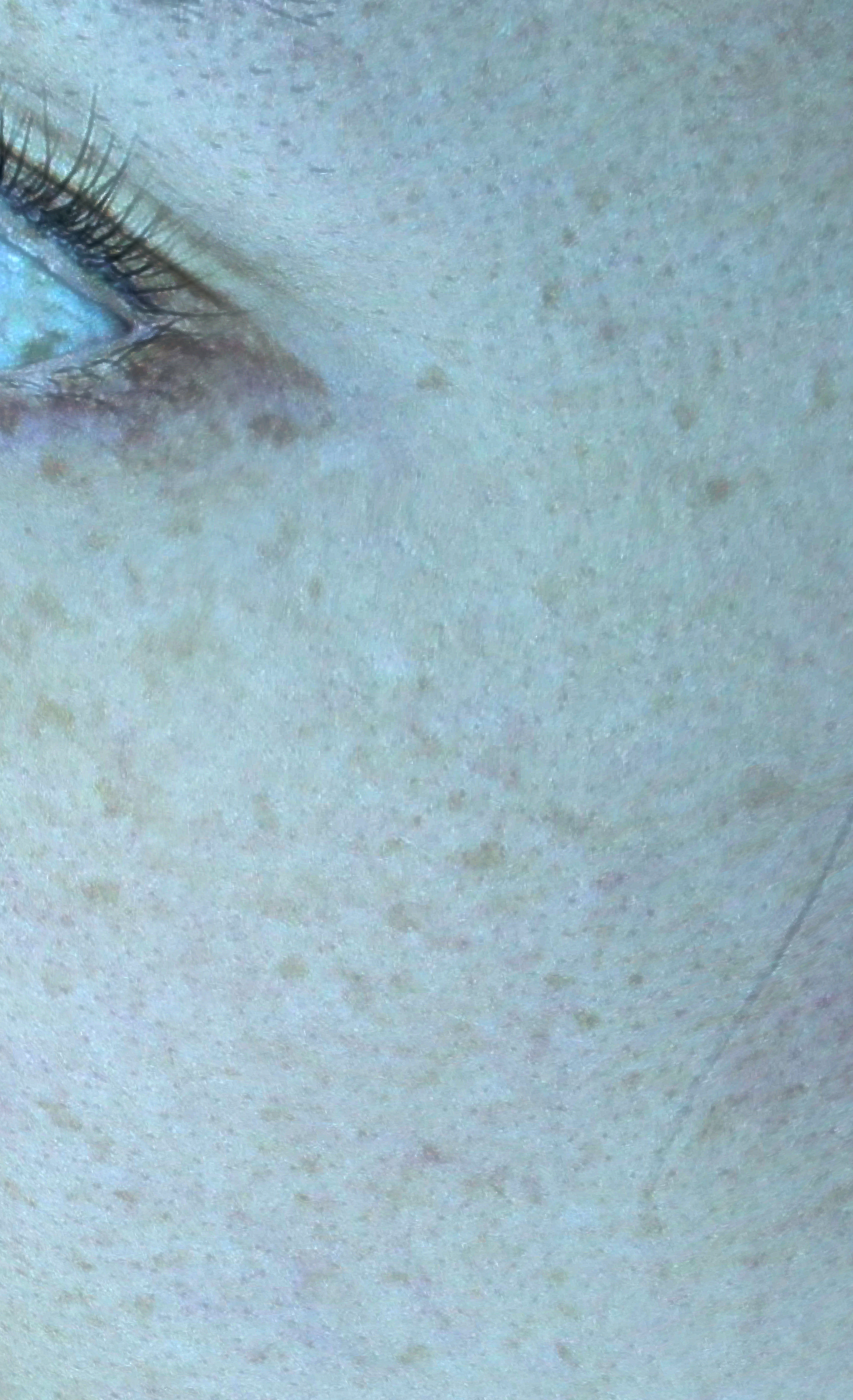
Rather than declaring the treatment finished after a single session, I tend to refine the direction while watching how the pigment responds. Especially with thin skin like that around the eyes, the course is often more stable when the approach is careful and divided into steps rather than forced. Because there are individual differences, the speed of response and the pattern of recovery vary from person to person.
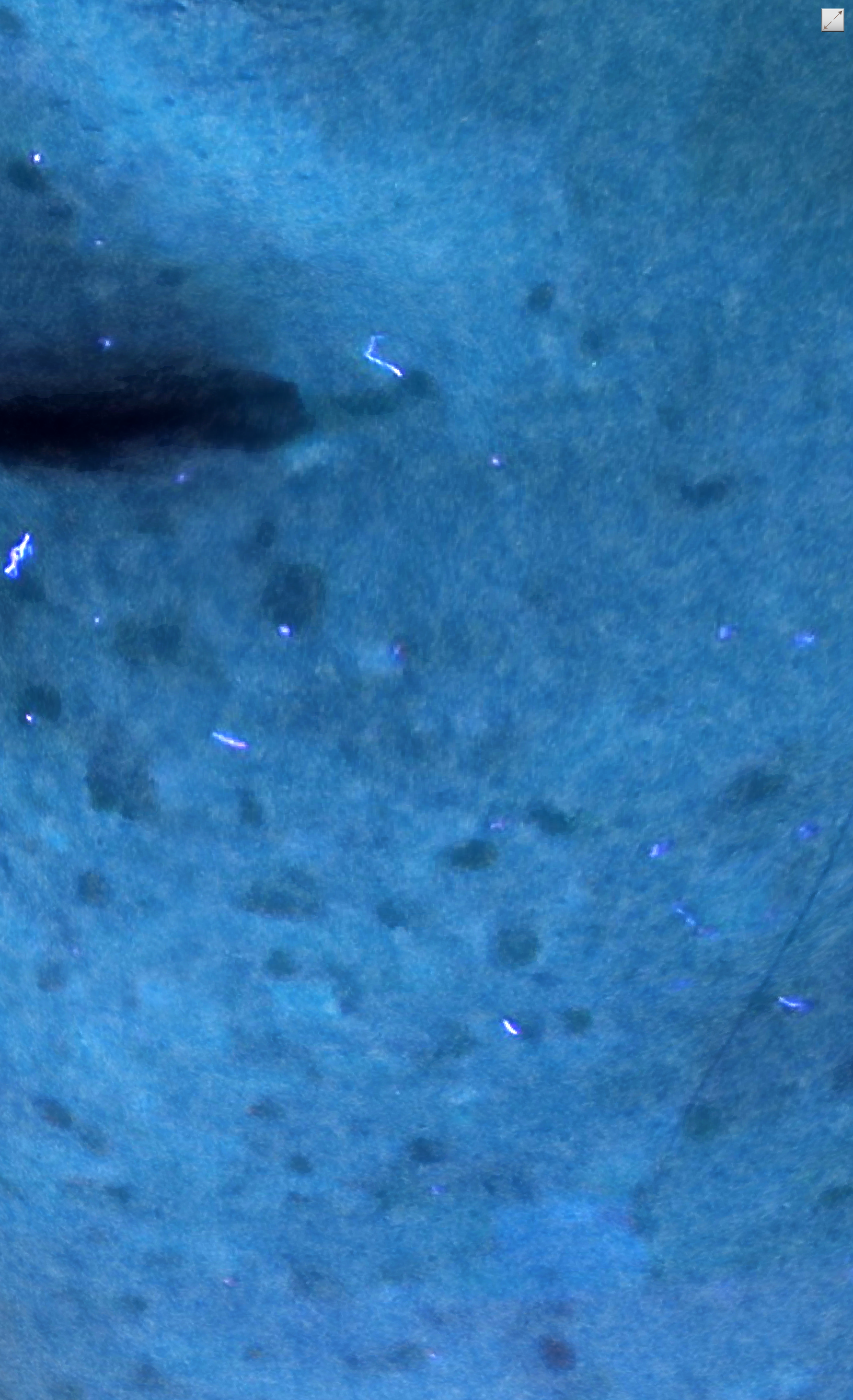
I have summarized why I consider the wavelength first, and why lentigo does not clear well with simple toning alone, in the records below.
- Why I weigh the wavelength first when choosing a laser for lentigo
- Why lentigo does not clear with simple toning
- A record of facing an eye-area lentigo that had recurred for more than 10 years
You can find an explanation of the Reepot laser itself in the Reepot laser guide.
Recovery care after removal matters even more
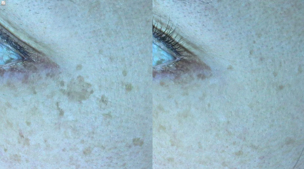
Right after the pigment has been treated, crusts or temporary color changes may appear. How you care for the skin during this period can determine the later course. In particular, UV exposure can re-stimulate the treated pigment, so during recovery I ask you to keep up sun protection and moisturizing consistently.
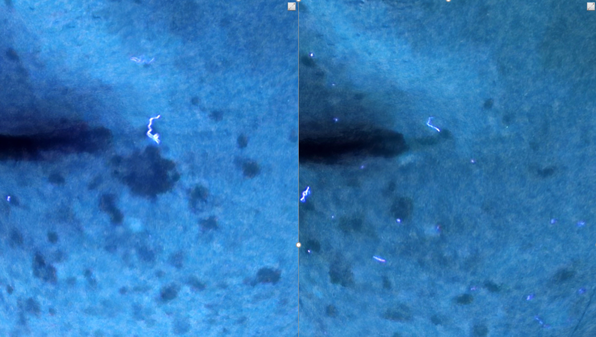
I have written in more detail about care after pigment removal in Why what comes after removing lentigo matters more. In the clinic I always emphasize that if recovery care is neglected, pigment that was carefully treated may appear to come back.
In the end, it starts again from diagnosis
To summarize:
- When I see brown pigment, I first determine whether it is lentigo or melasma.
- I set the wavelength and fluence according to the character and location of the pigment.
- In sensitive areas such as around the eyes, I do not force it and approach it in steps.
- I regard recovery care after removal as being as important as the procedure itself.
Because lentigo and melasma are treated in completely different ways, it is safer to decide on a direction only after a diagnosis by a board-certified dermatologist. Costs and the specific treatment plan are explained during the consultation after I have examined the lesion directly. If pigmentation around your eyes concerns you, feel free to start by asking about your condition below.
Consult via chatAuthor: Dr. Cho Mingyeol, Medical Director (board-certified dermatologist; Master Certification in Pigmentation, Acne, Rosacea and Hair from the Korean Dermatologists Association). You can find the full profile on the Medical Team page. First published 2024-12-16 / Last reviewed 2026-07-09
This article is intended for general information only; the appropriate treatment and outcomes may vary depending on each individual's condition.