

When I encounter pigmented lesions in the clinic, I think first of the principle by which a color is produced, rather than the color itself. This is because, even though it is the same melanin pigment, some lesions look deep black, some look faint brown, and others look unexpectedly bluish. Today I would like to borrow a bit of physics to explain how a dermatologist gauges the depth of pigment with the eye.
Like reading the depth of a lake from its ripples
We can roughly guess the depth of a lake just by looking at its ripples. The stretch where gentle ripples suddenly disappear and the surface becomes flat is usually where the water gets deeper. This is because the deeper the water, the longer the wavelength that appears on the surface.
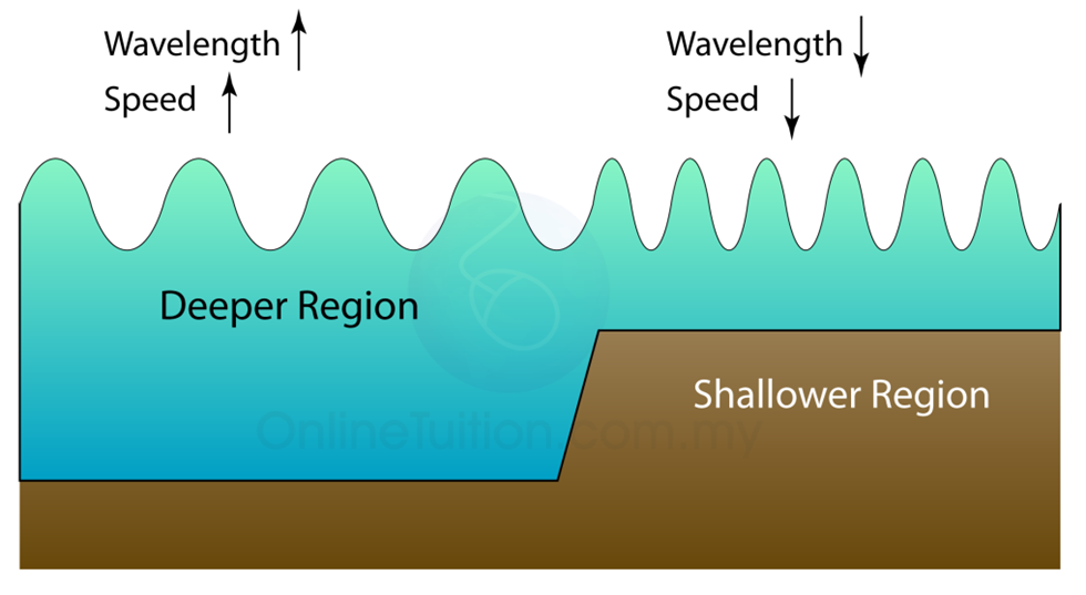
You may wonder why I am bringing up ripples in the middle of talking about pigment. It is because light, too, is an electromagnetic wave with a wavelength, and light passing through the skin can be understood in a way similar to ripples.
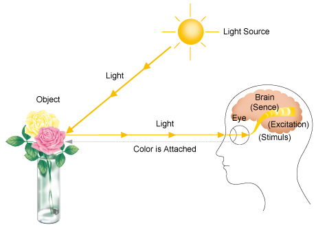
We see the light that is not absorbed as color
We perceive light and shade and color by detecting the light reflected from an object. Paradoxically, we perceive the color that an object does "not" absorb as that object's color.

If an object reflects all light, it looks white; if it absorbs all light and reflects none, it looks black. The color of the melanin made by melanocytes is originally brown (more precisely, it reflects brown). Yet actual pigmented lesions are observed in a variety of colors: black, dark brown, light brown, and sometimes even blue.
Where does this difference come from? To give the conclusion first, it is because of the location and density of the melanin pigment that the same pigment can look like a different color.
Depth and density determine color
External light penetrates into the skin, where it is absorbed or scattered, and color is perceived when the light that returns to our eyes is detected. Depending on where the melanin is and how densely packed it is, the nature of the returning light changes.
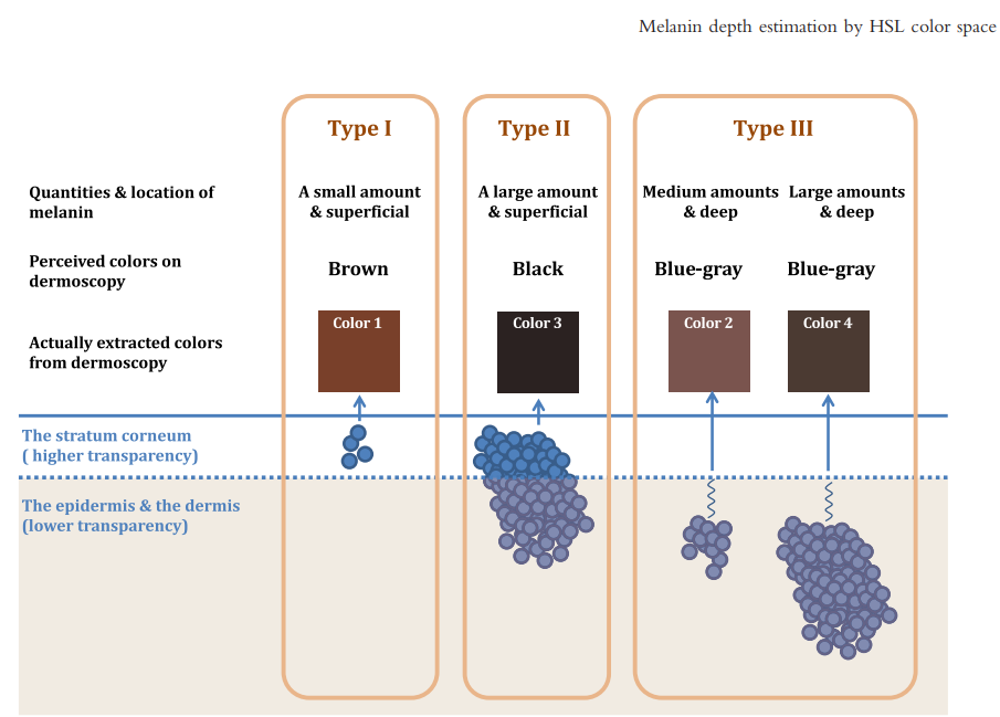
- When the pigment is at the skin surface and its density is high, both short- and long-wavelength light are absorbed by the melanin, and the lesion looks black.
- When the pigment is at the surface but its density is low, it looks brown.
- When the pigment lies deep, relatively short-wavelength light (in the blue range) scatters more within the tissue and returns to the eye, while longer-wavelength light is absorbed by the deep melanin, so the lesion looks blue.
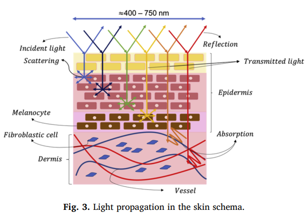
In summary, it can be divided as follows.
| Location of pigment | Density | Visible color |
|---|---|---|
| Epidermis (shallow) | High | Black |
| Epidermis (shallow) | Low | Brown |
| Dermis (deep) | Not relevant | Blue |
Knowing this principle helps you understand why color can come back even when only the surface melanin has faded. I have covered the perspective of looking at the dermal background as well in more detail in A story about pigment that requires reading both the epidermal and dermal background.
Color is not the identity of the pigment but the result of how the depth and density at which the pigment lies interact with light. That is why I see color as a clue, not a diagnosis.
Why I turn on the Wood's lamp when the naked eye is not enough
When pigments are mixed or it is hard to judge with the naked eye alone, I use a Wood's lamp (365 nm), which emits light of a shorter wavelength than visible light, to gauge the depth.
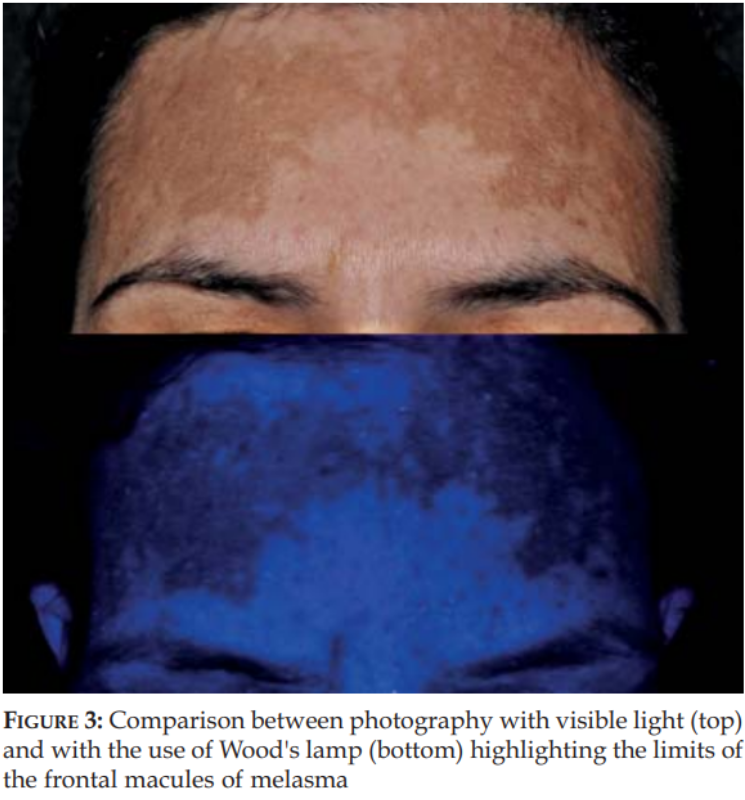
When the pigment lies deeper than the light of the Wood's lamp reaches, that is, when it is mainly dermal pigment, the wavelengths that enter the tissue pass through and scatter in the tissue above the pigment, and most of the light returns to the eye, so the lesion actually looks brighter.
Conversely, if it is epidermal pigment sitting shallowly, the surface pigment absorbs most of the light and very little returns, so it looks much darker. In other words, even for the same lesion, clues about depth emerge depending on how light is applied.
That is why the choice of wavelength and intensity differs
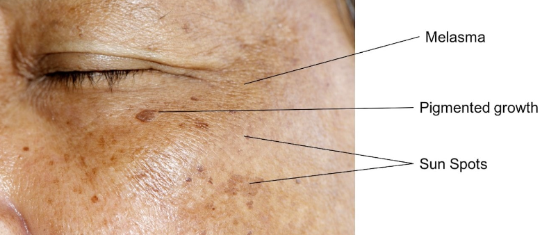
In actual practice, rather than appearing on their own, these lesions are more often found as several types mixed together on one person's face. Dermatologists use this principle to check the borders of vitiligo, or to read the depth of pigmented lesions and choose the appropriate laser wavelength, pulse width and intensity.
It may seem complicated, but as you observe and treat pigmented lesions over a long period, you build a sense that generally points you in the right direction. I have summarized my approach of dividing pigment into three types rather than lumping it together by appearance in Why I divide pigment that comes in as "age spots" into three types, and my judgment of considering wavelength first according to depth in Why I consider wavelength first in lentigo laser treatment. Why I recheck wavelength according to the season continues in Summer pigment treatment and wavelength.
That said, I do not diagnose a pigmentary disorder by the color and depth of the pigment alone. It must be judged by considering the age of onset, distribution, pattern, past history and more, which is why I can say that experience matters above all.

If you are concerned about a pigmentary condition, rather than judging for yourself based on color alone, having the depth and distribution examined together in the clinic will help set the direction. If you are curious which tests and approach would suit you, feel free to ask through Consult via chat.
References
- Sakai, Hiroshi, et al. "Estimating melanin location in the pigmented skin lesions by hue–saturation–lightness color space values of dermoscopic images." The Journal of Dermatology 44.5 (2017): 490-498.
- Handel, Ana Carolina, Luciane Donida Bartoli Miot, and Hélio Amante Miot. "Melasma: a clinical and epidemiological review." Anais brasileiros de dermatologia 89 (2014): 771-782.
- García, A. Madrid, and P. R. Horche. "Light source optimizing in a biphotonic vein finder device: Experimental and theoretical analysis." Results in Physics 11 (2018): 975-983.
Author: Dr. Cho Mingyeol, Medical Director (board-certified dermatologist; Master Certification in Pigmentation, Acne, Rosacea and Hair from the Korean Dermatologists Association). You can find the full profile on the Medical Team page. First published 2024-01-15 / Last reviewed 2026-07-09
This article is intended for general information only; the appropriate treatment and outcomes may vary depending on each individual's condition.