

When I consult with patients about lentigo in the clinic, I am often asked, "Why can't this be cleared neatly in one go?" I believe the answer lies in histology. Following my previous post summarizing pigmentary conditions easily confused with seborrheic keratosis, this time I would like to look at the histological background of lentigo.
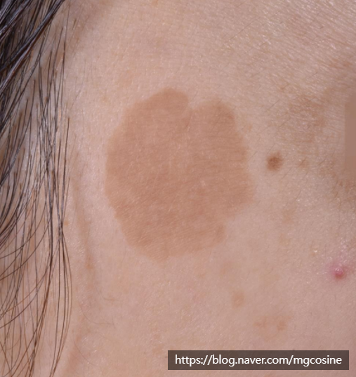
To treat lentigo well, it is better first to understand how the lesion is situated within the skin. So today I will go through the histological characteristics of solar lentigo one by one.
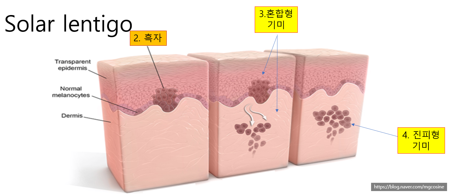
Why I start the lentigo story with histology rather than lasers
The histological characteristics of lentigo can be divided into five broad categories. First, let me summarize the big picture in a table. The content below is also the framework that runs through this entire post.
| Histological change | Why it makes treatment tricky |
|---|---|
| Melanocyte activation and elongation of dendrites | Melanin rises to the surface and the color darkens |
| Abnormal proliferation of keratinocytes | Melanosomes cannot be shed and accumulate |
| Elongation and descent of the rete ridges | Greater risk of damage to the upper dermis |
| Vascular proliferation | Possible vascular damage when strong energy is applied |
| Aging changes in the dermis | Hyperpigmentation and worsening of melasma appear more readily |
How do melanocytes make lentigo darker?
The melanocytes within a lentigo are activated by UV light and produce excessive melanin pigment. Up to this point, it is the course you would expect of a pigmentary disorder.
The problem is what comes next. When melanocytes are excessively activated, their dendrites grow longer, and the further the melanosomes they produce rise toward the epidermal surface, the blacker and darker the lentigo becomes. Looking at three-dimensional reconstructions from electron microscopy, you can see that the melanocyte dendrites of senile lentigo (bottom d, e, f) are longer and more branched than those of normal skin (top a, b, c).
The traces left by keratinocyte proliferation
Strong UV exposure causes DNA damage, and this damage also affects the epidermal cells. As a result, abnormal proliferation of keratinocytes, the cells that make up most of the epidermal layer, appears in lentigo areas.
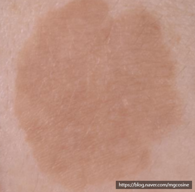
When keratinocytes increase, melanosomes cannot be shed normally and simply accumulate. This is the background to why lentigo darkens. Lentigo in which the epidermis itself has been altered in this way is not easy to remove merely by repeating high-power toning. I touched on a similar context in my post on why laser toning did not respond as expected.
If, having read this far, you are wondering which side your own lesion is closer to, feel free to ask via Consult via chat now.
The problem that arises when rete ridges descend into the dermis
In solar lentigo, the rete ridges elongate from the basal layer of the epidermis, increasing the interaction between melanocytes and keratinocytes. This promotes the transfer of melanosomes and makes the lentigo stand out even more.
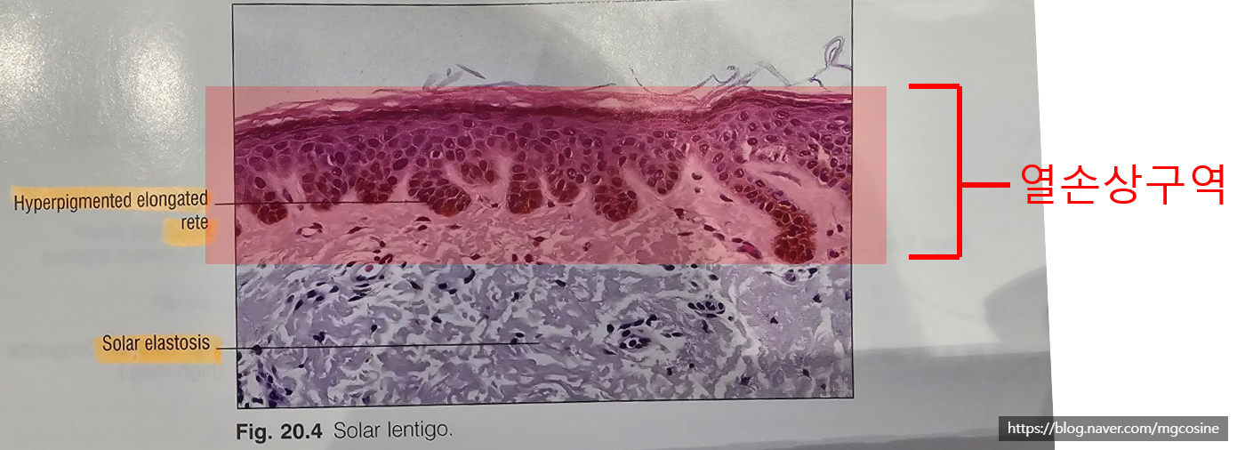
On top of this, the rete ridges extend downward and settle deeper, into the upper dermis. So when lentigo is treated, the possibility of damaging the upper dermis rises as well. This point is clinically important.
When the upper dermis is disturbed, phenomena such as post-inflammatory hyperpigmentation or worsening of melasma can appear more readily. This is exactly the histological factor that makes lentigo treatment difficult.
Why are blood vessels also increased in lentigo?
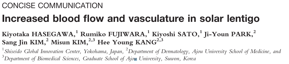
As seen in the paper above, lentigo also shows findings of vascular proliferation. Because lentigo has marked epidermal changes, in theory an approach that clears the epidermis at once with strong energy is needed. But doing so can bring the problems listed below.
- The likelihood of damaging the proliferated blood vessels increases
- Vascular damage can lead to post-inflammatory hyperpigmentation
- As a result, it can sometimes lead to melasma worsening as well
Treating it strongly raises concerns about the blood vessels and dermis, while treating it gently leaves the epidermal changes behind; this kind of dilemma arises often. That is why lentigo treatment is by no means simple.
Lentigo ultimately meets aging
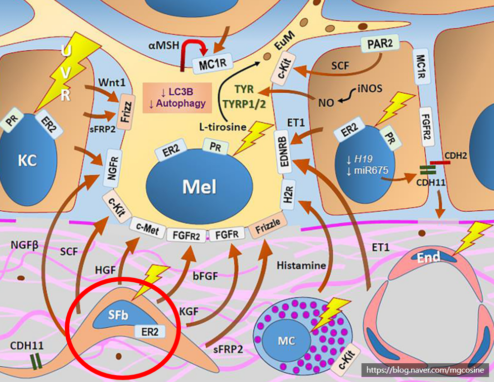
These days there are many reports that melasma is associated with aging. Lentigo lesions are also deeply intertwined with aging. The fact alone that lentigo lesions are almost never seen in teenagers shows this.
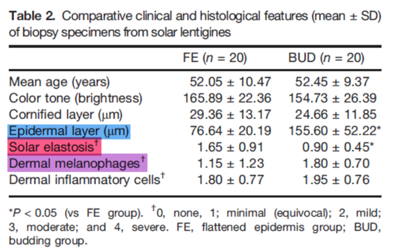
In lentigo too, signs of dermal aging are observed, namely senescent fibroblasts (SFb) and solar elastosis. If treatment proceeds in the wrong direction against this background, hyperpigmentation and worsening of melasma can become more severe, and the lentigo lesion may actually darken. This also connects with the early management of post-inflammatory hyperpigmentation, which is often left behind when lesions are treated the wrong way. My honest impression from the clinic is that lentigo treatment is just as tricky as melasma.
The direction for lentigo treatment that histology points to
In summary, lentigo is a pigmentary disorder arising from several overlapping factors: abnormal proliferation and structural alteration of melanocytes and keratinocytes, vascular proliferation, and dermal aging. Because there is not just one cause, it is hard to push through with a single setting.
So when I look at a lentigo, I try to read the depth of the lesion and the condition of the epidermis, blood vessels and dermis together before deciding on a direction. Actual laser selection and approach are covered more concretely in the lentigo laser treatment post and in the introduction to the Reepot laser used at our clinic. The suitable method differs from lesion to lesion, so I always mention that there are individual differences.
Consult via chat
Author: Dr. Cho Mingyeol, Medical Director (board-certified dermatologist; Master Certification in Pigmentation, Acne, Rosacea and Hair from the Korean Dermatologists Association). You can find the full profile on the Medical Team page. First published 2024-07-03 / Last reviewed 2026-07-09
This article is intended for general information only; the appropriate treatment and outcomes may vary depending on each individual's condition.