

Hello. I am Cho Mingyeol, a dermatologist at OARO Dermatology. Today I would like to record a case of multiple lentigines that I encountered again in the clinic, including my decision-making process.
Lentigo on the cheekbone for 4 years: how did she present?
A woman in her mid-30s came in with lentigo on her right front cheek and cheekbone area as her chief complaint. She said the pigment had first become noticeable about 4 years earlier. In the photos, a pattern of solar lentigo with relatively distinct borders can be observed.
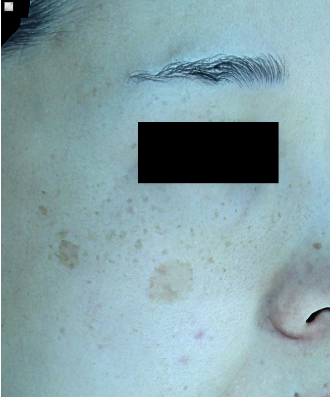
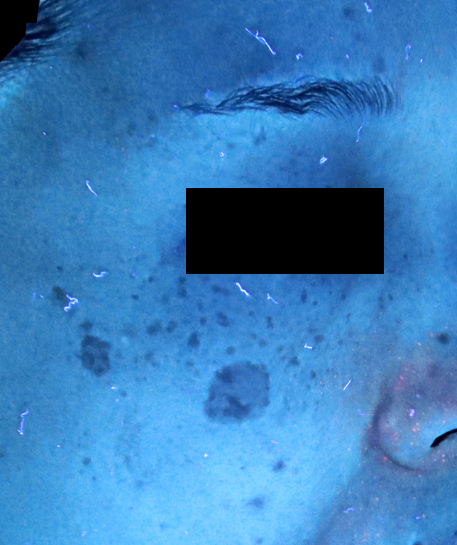
Solar lentigo is a pigmentary disorder that develops as long-term UV exposure accumulates and melanocytes in the basal layer of the epidermis partially increase. Many people complain that as it grows, even makeup cannot fully cover it, so it keeps bothering them in daily life. This patient, too, had long struggled with that discomfort before knocking on the clinic door. I have separately explained how lentigo becomes established histologically in Why lentigo forms, explained through histology.
Why I approached this pigment with the Reepot laser
There is one principle I always keep in mind in pigment treatment.
Lightening pigment depends not on how strong an output you use, but on which pigment, at which depth, you target.
This lesion was a distinct lentigo spread widely across the epidermis, on skin where the surrounding redness and vascular reactions also needed to be considered. So I approached it with the Reepot laser, which is designed to deliver energy to pigmented lesions while momentarily calming the superficial blood vessels. The goal is not strong output itself; the key is to deliver precisely as much energy as the targeted pigment needs.

I have covered the specific principles and settings for approaching lentigo with the Reepot laser in more depth in Part two on treating lentigo with lasers, so if you are curious about the principles, you may want to read it as well.
Right after treatment, how did the pigment change?
Let's follow the course after treatment.
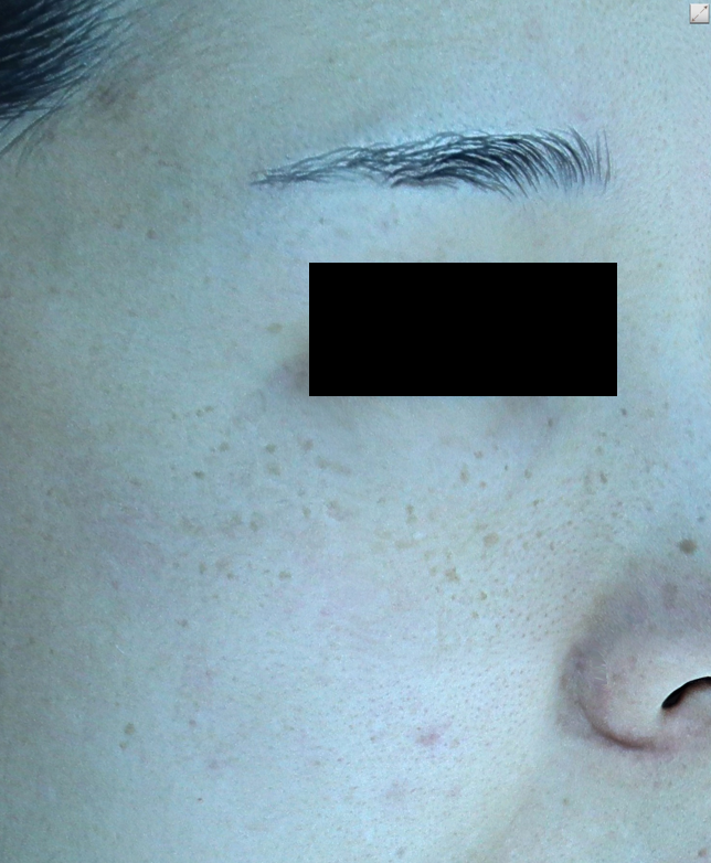
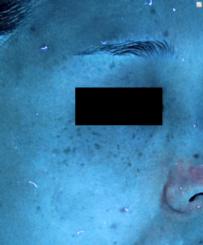
Though there are individual differences, the pigment that had spread widely had faded noticeably. Compared with her first visit, the borders of the lesion had become considerably blurred. Of course, I cannot say that this result appears in exactly the same way for every lentigo. The course inevitably differs depending on skin thickness, the depth of the pigment, and sun protection habits.
Placing before and after side by side
Let's compare the before and after photos under the same conditions.
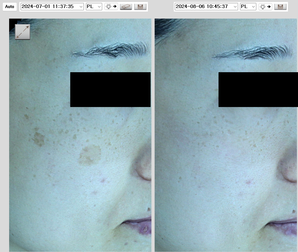
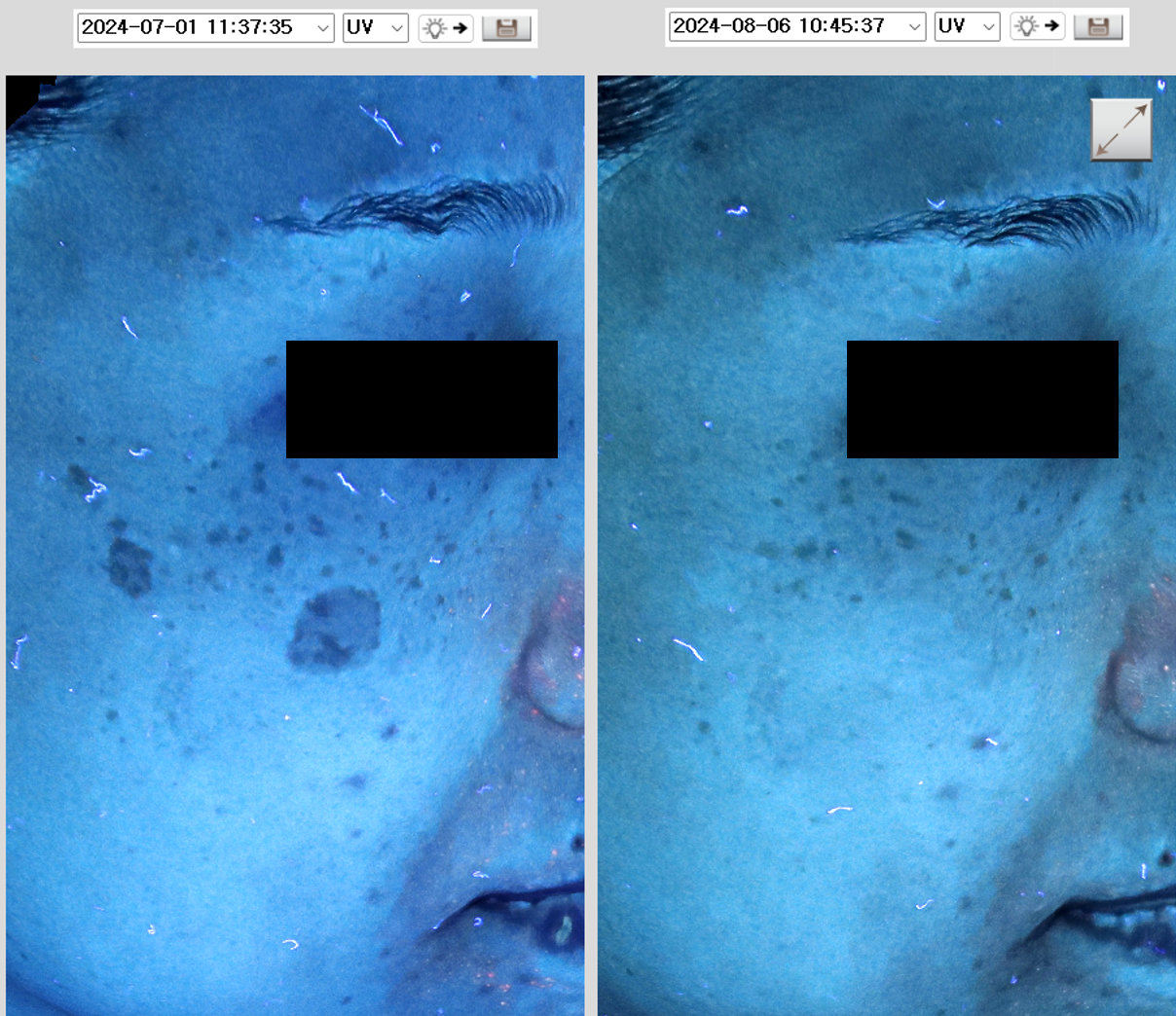
When I look at before and after photos, the points I actually check in the clinic are as follows.
- Have the borders of the lesion blurred, or has only the color faded?
- Has the difference in tone from the surrounding normal skin decreased?
- Has recovery progressed stably, without redness or signs of irritation?
Only when these three improve together do I judge the course to be satisfactory.
Where the lentigo was removed: the moment the DuoDERM comes off
After the laser, the lesion is covered with a thin dressing (DuoDERM) to aid recovery. I have captured the dressing being removed.
In my view, in lentigo treatment, the management of the recovery period after removal determines the final result more than the moment the laser is fired. If crust care, sun protection, or soothing care goes off track, pigment that had faded can come back up. I have written in detail about what direction of care I ask for after treatment in What I ask of you during recovery after lentigo removal.
What it means that not everything is erased at once
After her lentigo faded following treatment, this patient said she could now see other pigment that had been hidden behind the lentigo. This is not a failure but a natural sequence often encountered in pigment treatment. Only once the darkest and widest pigment is cleared do the fainter pigments lying beneath finally come into view.
That is why, at the first consultation, I always tell people this: it is difficult to guarantee that all pigment will disappear with a single treatment, and it needs to be approached in stages, by type of pigment. Lentigo in particular is easily confused with seborrheic keratosis or other pigmentary disorders, so the differential diagnosis that decides what to address first comes first.
| Category | Commonly seen features | Approach I take in the clinic |
|---|---|---|
| Solar lentigo | Distinct brown spots on sun-exposed areas | Step by step, with a laser targeting the pigment |
| Seborrheic keratosis | Keratotic lesions with a slightly raised surface | A separate approach matched to lesion thickness |
| Melasma | Blurred borders, spreading bilaterally | Care that also considers the dermal background |
I have explained why pigment is hard to classify by eye alone in Pigmentary disorders easily confused with age spots. A dermatologist with broad experience in pigment treatment will help you set a direction, so if you are wondering what kind of pigment is bothering you now, please feel free to contact us.
Consult via chatThe photos used in this post were taken and published with the patient's consent, and unauthorized use is prohibited.
Author: Dr. Cho Mingyeol, Medical Director (board-certified dermatologist; Master Certification in Pigmentation, Acne, Rosacea and Hair from the Korean Dermatologists Association). You can find the full profile on the Medical Team page. First published 2024-09-23 / Last reviewed 2026-07-09
This article is intended for general information only; the appropriate treatment and outcomes may vary depending on each individual's condition.